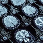
What are MRIs?
Most people with MS are very aware of MRI or Magnetic Resonance Imaging, often having regular MRIs to diagnosis and monitor their MS. Most people don’t love them, but MRIs are a marvel of modern medicine, allowing us to visualise what is going on in the human body and a rare opportunity to peer beneath the skin.
MRIs use strong magnets and radiofrequency pulses to generate signals from the body. These signals are in turn detected by the machine and are processed by a computer to create pictures of the inside of our bodies.
How does MRIs work?
The human body is largely made of water molecules. Each water molecule is comprised of two hydrogen and one oxygen atom and due to the chemical properties of these atoms, water molecules (and other molecules in the human body) act like miniature weak bar magnets with a north and south magnetic pole. Normally these molecules are just randomly jumbled up in the body. However, in an MRI machine the human body is placed in a huge magnet which causes the water molecules to all align up in one direction. The MRI machine then changes the direction of the pulses causing the molecules in the human body to flip orientation. How quickly they change depends on the chemical composition of each molecule and on what other chemicals are around it. The MRI machine then detects these billions of minute changes and how long they last and builds up an image of the composition of the body.
The magnetic field is created by passing electricity through coils within the machine, which can cause the coils to vibrate, resulting in a knocking sound inside the scanner. These noises can be loud and concerning but are completely normal.
MRIs and MS
MRI can be used to look at most types of tissues and body parts and can diagnose a variety of conditions. In MS, MRIs are used to look at the brain and spinal cord (the central nervous system).
By detecting the subtle differences in body composition, MRIs can identify areas of inflammation and damage within the central nervous system. In MS these are the result of the immune system attacking and damaging the insulating layer of nerve cells, known as myelin.
Myelin helps nerves to conduct their electrical signals very quickly, as well as acting as a protective layer and supplying nourishment to the cells beneath it. Loss of myelin results in failure of nerve signals and can ultimately result in the death of nerve cells.
MRIs can help detect these areas of inflammation and can also detect the scarred patches where cells have died. MRIs however, may also be able to detect other more subtle effects of MS on the brain, with studies over the last few years revealing another important emerging measure and that is changes in brain size or volume known as brain atrophy.
Areas of inflammation and damage in the brain or on the spinal cord leave marks on the MRI images that look like spots and are referred to as lesions.
The positions of these spots can give an indication of symptoms, for example lesions on the spinal cord, can indicate numbness or loss of movement of some of the limbs. However, often spots cannot be linked to specific symptoms and may be referred to as asymptomatic or ‘silent’. It is thought that as many as 9 in 10 lesions do not result in a bout of symptoms or a relapse. Stopping new lesions is considered to be the main goal of treatment, however in some cases this doesn’t prevent the continual progression of the disease.
Decoding types of MRI images in MS
The MRI field is awash with acronyms and jargon which refer to the different types of pictures (or sequences). These are generated by changing the strength and duration of the magnetic and radio pulses which cause the magnetic molecules in the body to change their behaviour. The different sequences are a bit like a photographer using different lighting and changing the aperture and lens to capture perfect photos.
Here are some of the most common types of MRIs in MS;
- T2/Flair images. When radiologists carry out a T2/Flair sequence MS lesions stand out as bright spots which are often referred to as hyper intensities. These show spots of both new and old damage. In MS these tend to cluster deep in the brain and these pictures are used to determine how active the disease currently is.
- T1 with contrast images. To help identify active lesions, the radiologist may use a contrast agent or a chemical which shows up brightly on an MRI, a commonly used agent is a chemical known as Gadolinium (GAD) which is injected into the vein prior to the MRI. GAD does not normally en¬ter the brain or spinal cord however when the blood ves¬sels are ‘leaky’ due to inflammation it can leach out of the blood vessels and show up as an area of brightness in the MRI image. This indicates an active lesion. Typically, these are only visible for a couple of weeks to a couple of months and often then revert to a T2/Flair scar.
- T1. These images are carried out in a similar way to the previous sequence but without a chemical agent. On these images, areas where there has been damage to the myelin coating and nerve cells appear as black holes.
Reading MRIs is a complicated endeavour, and not all lesion like marks appearing on MRIs are due to MS. Things like injury, infection, and blood vessel blockages can all also show up as spots on an MRI.
Traditionally neurologists have used the presence of new lesions in the MRIs of a person with MS as a sign that a change in medication may be needed, but more sensitive measures are also now being included in the assessment of MRI images.
Other MRI measures
While lesions have been the critical measure considered on the MRI images, more recent research has revealed that brain atrophy, or shrinkage in brain volume, may be a more accurate measure which potentially correlates more closely with disease progression and disabilities than lesions and relapses.
Brain atrophy or brain shrinkage is a normal process of ageing, but it can be accelerated in people with MS. On average we lose between 0.5% and 1% of our brain volume per year and this varies greatly from person to person, however for people with MS, shrinkage can be slightly higher.
Measuring the rate of brain atrophy is slightly challenging, it requires regular high-quality MRI scans that are done in a comparable way every time. Changes in volume are small and are therefore difficult to detect on images which were taken on different MRI machines and by different radiologists.
Recent research into both relapsing and progressive MS has shown that people with more brain atrophy have a poorer outcome and studies have shown that this is independent of the number of lesions.
Research also shows that the spinal cord can shrink too, and that this is more marked in primary progressive MS than in relapsing remitting MS. Additional research has also shown that atrophy of certain subsections of the brain may be more important than overall atrophy. Importantly, studies have also shown that some MS medications can slow the rate of atrophy bringing it more in line with normal ageing.
Brain volume measures are becoming more accessible in clinics but further work is required to establish a unified way of taking MRI pictures and to assess them to accurately measure atrophy. MS Research Australia funded researchers such as Dr Heidi Beadnall, Brain and Mind Centre, NSW have been working on sensitive and automated tools to help improve the routine detection of atrophy in clinical practice.
The current goal of therapies is now to achieve no evidence of disease activity (known by the acronym NEDA). Traditionally, treatment of relapsing remitting MS has focused solely on reducing the number of relapses, however given the activity seen on MRI, we know there is additional activity other than relapses which we need to treat to achieve NEDA, and that includes lesions seen on MRIs and potentially also atrophy.
The Future of MRI
The field of MRIs has exploded. It was only in the early 1980’s that saw the introduction of MRIs and since then we have seen the rapid evolution of this technology. Over its relatively short existence, MRI has become the imaging technique of choice for the study and clinical evaluation of MS and other diseases, with more than 60 million clinical MRI scans performed annually on over 25,000 MRI systems world-wide.
The introduction of more powerful machines has given even more detailed vision into the human brain and has brought about the possibility of measuring brain and spinal cord shrinkage. Future advancements will only further enhance this and allow us to peer into the brain and gather even more information on health and disease. Combining detailed imaging with big data on clinical outcomes, also provides potential for artificial intelligence computing to sift through hundreds of scans and detect subtle changes even earlier and more accurately, accelerating and enhancing the diagnosis of MS and management of MS.